| Synonyms | A170,DMRV,EBI 3 associated protein of 60 kDa,EBI 3 associated protein p60,EBI3 associated protein of 60 kDa,EBI3 associated protein p60,EBI3-associated protein of 60 kDa,EBIAP,FTDALS3,MGC127197,ORCA,OSF-6,Osi,OSIL,Oxidative stress induced like,p60,p62,p62B,Paget disease of bone 3,PDB 3,PDB3,Phosphotyrosine independent ligand for the Lck SH2 domain of 62 kDa,Phosphotyrosine independent ligand for the Lck SH2 domain p62,Phosphotyrosine-independent ligand for the Lck SH2 domain of 62 kDa,PKC-zeta-interacting protein,Protein kinase C-zeta-interacting protein,Sequestosome 1,Sequestosome-1,SQSTM 1,SQSTM,Sqstm1,STAP,STONE14,Ubiquitin binding protein p62,Ubiquitin-binding protein p62,ZIP 3,ZIP,ZIP3 |
| Swissprot | Q13501 |
| Source | Rabbit |
| Reactivity | Human |
| Immunogen | Synthetic peptide of human SQSTM1 |
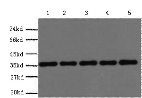
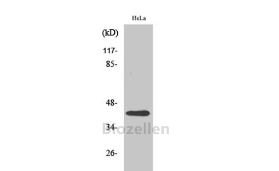
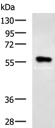
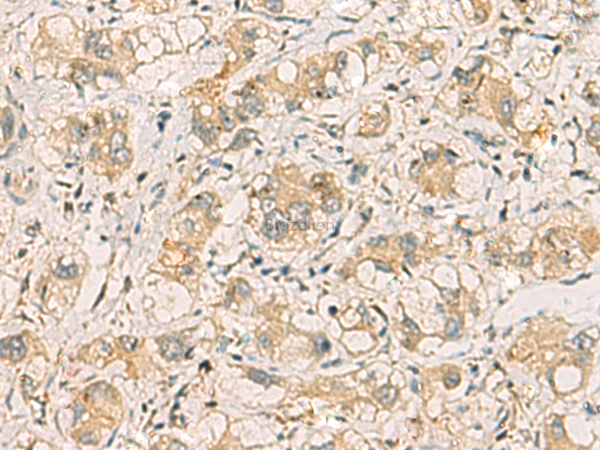
| Application | WB,IHC,ELISA |
| Recommended dilution | WB 1:500-1:2000, IHC 1:50-1:100, ELISA 1:5000-1:10000 |
| Concentration | 1.14mg/mL |
| Clonality | Polyclonal |
| Cellular localization | Cytoplasm. Late endosome. Nucleus. Sarcomere (By similarity). In cardiac muscles localizes to the sarcomeric band (By similarity). Localizes to late endosomes. May also localize to the nucleus. Accumulates in neurofibrillary tangles and in Lewy bodies of neurons from individuals with Alzheimer and Parkinson disease respectively. Enriched in Rosenthal fibers of pilocytic astrocytoma. In liver cells, accumulates in Mallory bodies associated with alcoholic hepatitis, Wilson disease, indian childhood cirrhosis and in hyaline bodies associated with hepatocellular carcinoma. |
| Isotype | IgG |
| Purification | Antigen affinity purification |
| Conjugation | Unconjugated |
| Storage instructions | Store at -20℃. Avoid freeze / thaw cycles. |
| Storage buffer | PBS with 0.05% NaN3 and 40% Glycerol,pH7.4 |
| Background | Sequestosome 1 (SQSTM1/p62) is a multifunctional adaptor protein implicated in selective autophagy,cell signaling pathways,and tumorigenesis. p62 has been implicated in shuttling ubiquitinated and sometimes aggregated proteins for autophagic degradation. As a autophagy-specific substrate,p62 is degraded during the autophagic process,which makes intracellular level of p62 as a marker for autophagy flux. p62 is at the cross-roads of several signaling pathways including Ras/ Raf/ MAPK and NFκB and plays important role in cancer. p62 is a component of inclusion bodies/ protein aggregates found in human diseases,including Huntington's disease,Alzheimer's disease,Parkinson's disease in the brain,and nephropathic cystinosis in kidney (22074114,22860231,22714671). The molecular weight of p62 is predicted as 48/ 38 kDa,while western blot analyses using this antibody demonstrate the major band around 60-62 kDa in various tissues. |